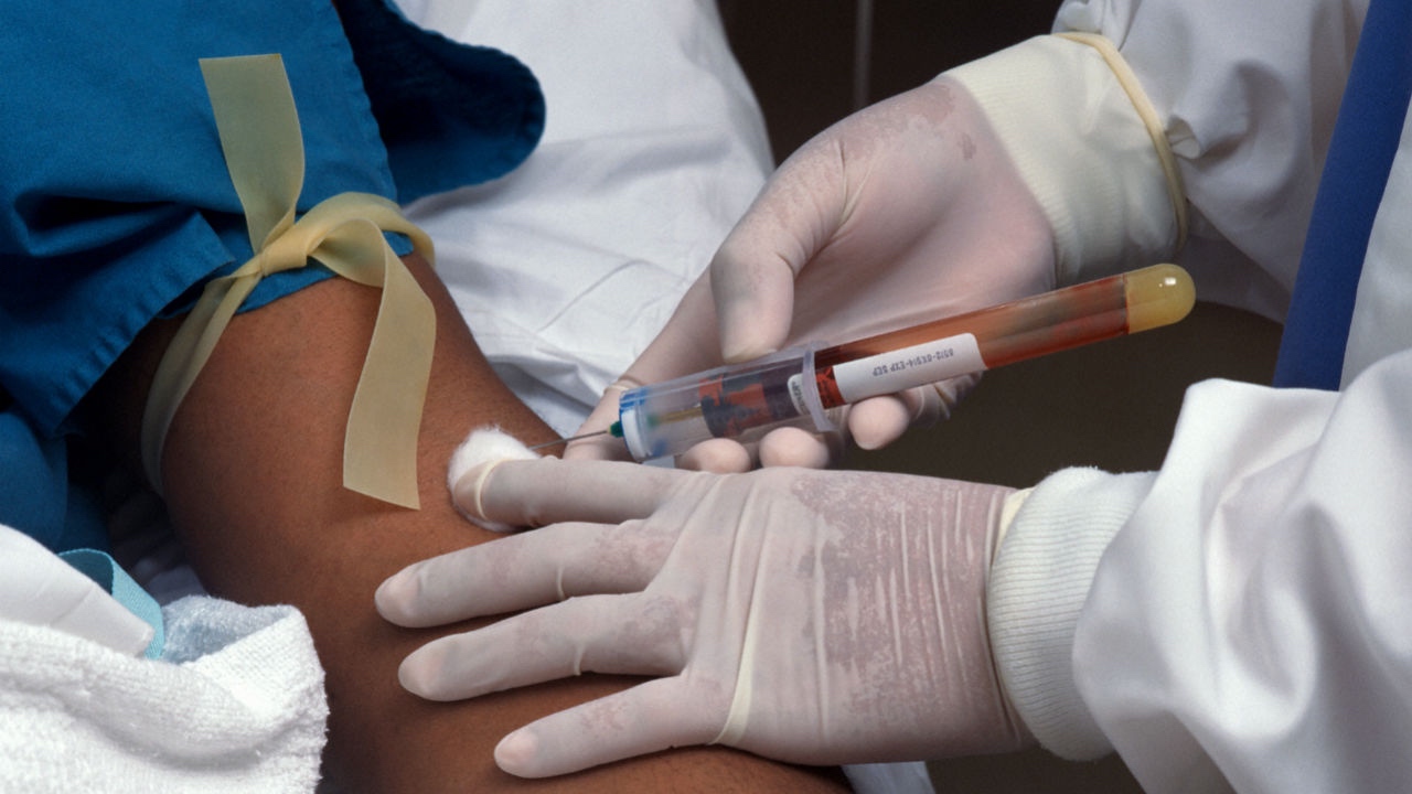
Blood Monitoring
Full blood Count:
Anaemia may be due to chronic disease but be careful to consider blood loss from gastric irritation secondary to NSAIDs or other causes of anaemia.
White cells: possible changes include neutrophilia in septic arthritis, eosinophilia in polyarteritis nodosa, neutropenia in Felty’s syndrome and leukopenia in SLE.
Platelets may be increased in RA and may be decreased in SLE.
ESR and CRP indicators of inflammatory activity.
Uric acid: may be raised in gout.
Renal function: may be renal dysfunction in chronic disease such as gout or connective tissue disorders.
Autoantibodies: rheumatoid factor may support the diagnosis of RA. An antibody to a substance called cyclic citrullinated peptide (CCP) has been found to be more specific than rheumatoid factor in RA and may be more sensitive in erosive disease.
Antinuclear antibodies may suggest SLE or other connective tissue disorders.
Human leukocyte antigen (HLA) B27: increased positivity in ankylosing spondylitis and other spondyloarthropathies.
Serology (eg, HIV), may be appropriate.
Other investigations
Urine: proteinuria may be due to nephrotic syndrome associated with connective tissue disease.
Synovial fluid:
White cell count raised in infection.
Gram stain (tuberculosis), culture and sensitivities.
Crystal identification: urate, calcium pyrophosphate.
Imaging:
X-rays: may show distinctive changes, such as in RA, and osteoarthritis. CXR may be indicated for lung involvement in RA, SLE, vasculitis and tuberculosis.
Ultrasound: soft tissue abnormalities – eg, synovial cysts.
CT scan, MRI: much greater information of bone, joint and soft tissue.
Arthroscopy:
Direct view of joint and synovial fluid.
Potential for biopsy and therapeutic procedures.

